Lab Alumni
- Nico Tenorio (Published!) | 2023-2024
- Victoria Colling (Published!) | 2022-2024
- Keaton Silver (Published!) | 2022-2024
- Audi Fineran (Published!) | 2022-2023
- Aidan Quinn | 2022-2023
- Dylan Poch: PhD student, Yale University (Published!) | 2019-2022
- Marissa Allen: PhD student, Marquette University | 2020-2022
- Mary Quansah (Published!) | 2021-2022
- Chris Chamblee: PhD student, Texas A&M | 2020-2022
- Andrew Smith: MD student, Kansas University (Published!) | 2019-2021
- Dr. Tyler Sodia: Particle Scientist, VitriVax; PhD, Colorado School of Mines (Published!) | 2018-2020
- Lindsey Armstrong: Lab technician, Creighton University | 2018-2020
- Austin Haider: PhD student, Denver University | 2018-2020
- Marcos Maldonado: (Published!) | 2016-2019
- Anika James: PhD student, Kansas University | 2018-2019
- Marlea Kudlauskas: Cardiac ICU Nurse | 2018-2018
- Anna Nguyen: PhD student, University of California Santa Barbara (Published!) | 2017-2018
- Lisa Fetter: PhD student, University of California Santa Barbara (Published!) | 2014-2018
- Derek Clark: Pharmacy Technician | 2017-2018
- Dr. Ilia Mazin: PhD, the Ohio State University | 2016-2017
- Dr. Nazar Dubchak: Anesthesiology resident, Walter Reed | 2016-2017
- Jena Jacobs: DO medical student, A.T. Still University | 2015-2017
- Dr. Jessica (Daniel) Watson: MD, University of Florida (Published!) | 2015-2016
- Susan Jett: Biobank Manager, Thermo Fisher (Published!) | 2015-2016
- Aviva Bulow: Software engineer, Sigray | 2014-2016
- Dr. Ryan Warren: Spectrum Health Lakewood, Michigan | 2015-2015
- Dr. Tiffany (Ashbaugh) Tesmer: DO, Midwestern University College of Osteopathic Medicine | 2015-2015
- Michael McCoy | 2015-2015
- Ebony Miller: Certified Nursing Assistant at University of Colorado Health Sciences Center | 2015-2015
- Laura Roon: PRA at Skaggs School of Pharmacy (Published!) | 2014-2015
- Jonathan Richards: Freelance Tutor (Published!) | 2013-2015
- Becky Addison: GIS Software Developer at USGS | 2013-2014
- Jeremy O’Brien: Canning Line Operator | 2013-2014
- Travis Ingraham: Director of Extraction and Quality Assurance for GCH USA | 2013-2014
- Sarai Graves: Provider Relations at United Health Group | 2013-2014
- Kathryn (Norquest) Vang: M.S.; PRA, National Jewish, Colorado (Published!) | 2012-2014
- Dr. Stephen Schaffner: DO, Rocky Vista Medical School (Published!) | 2012-2014
- Kyra Brandt: 1st Lieutenant Signal Officer, US Army | 2012-2014
- Elina (Baravik) Wegner: Oncology R&D at Invitae (Published!) | 2012-2014
- Yerelsy Reyna: Surgeon’s Assistant at Children’s Hospital (Published!) | 2012-2014
- Dr. Josh Sowick: MD, Rosalind Franklin University of Medicine and Science | 2012-2014
- Jody Stephens: Group Lead Software Testing at ArcherDx (Published!) | 2011-2014
- Dr. Ryan Masterson: Resident doctor, Johns Hopkins (Published!) | 2012-2013
- Mason Preusser: Consultant at D-A Lubricant | 2012-2012
- Dr. Tonya Santaus: PhD, University of Maryland Baltimore County | 2012-2012
- Amanda Faux | 2012-2012
- Matthew Stoddard: Scientific Informatics Consultant at Accenture | 2011-2012
- Morgan Miller: Digital Strategy Director at DMA Digital | 2011-2012
Nico Tenorio
Lab Member 2023-2024
Published
Victoria Colling

Lab Member 2022-2024
Published
Keaton Silver
Lab Member 2022-2024
Published
Audi Fineran

Lab Member 2022-2023
Published!
Aidan Quinn

Dylan Poch
Lab Member 2019-2022
Current: PhD student, Yale University
Published!
Marissa Allen

Lab Member 2020-2022
Current: PhD student, Marquette University
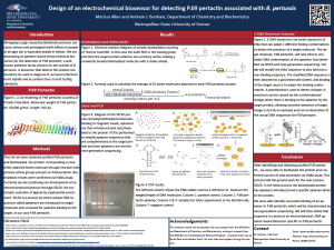
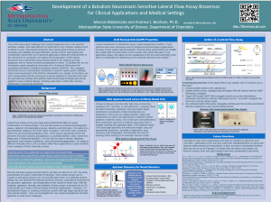
Whooping cough caused by Bordetella pertussis can cause serious and prolonged health effects, particularly in infants. Quick and accurate diagnosis is essential in treating the infection. Early treatment can significantly reduce the duration of illness in the patient and lead to a milder case of illness overall. The current standard for testing set forth by the CDC is invasive, requiring nasopharyngeal swabs. Most medical facilities are not equipped to then process the sample collected, and it is instead sent out for analysis adding to the length of time required to get a diagnosis for the patient. Common laboratory tests for the detection of B. pertussis are PCR and culture, but each present unique challenges. The purpose of our research is to design and develop a rapid electrochemical biosensor that can detect P.69 pertactin. P.69 pertactin is a surface protein used by B. pertussis to bind to mammalian cells. A biosensor that can detect P.69 pertactin would improve several major issues associated with the current testing standards including size of the patient sample needed, time to get results, cost of testing and shipping, and accuracy of tests. The B. pertussis biosensor will be developed through the SELEX process and will create a specific oligonucleotide probe for P.69 pertactin . This probe will then be adapted into an electrochemical DNA-based biosensor. This biosensor would require a much smaller sample from the patient and give almost immediate results to the physician overseeing the case.
Mary Quansah
Lab Member 2021-2022
Published!
Chris Chamblee

Lab Member 2020-2022
Current: PhD student, Texas A&M
Andrew Smith
Lab Member 2019-2021
Current: MD Student, Kansas University
Published!
Dr. Tyler Sodia
Lab Member 2018-2020
Current: Particle Scientist, VitriVax; PhD, Colorado School of Mines
Published!
Lindsay Armstrong
Austin Haider
Current: PhD student, Denver University
Lab Member 2018-2019
Project: Algorithmic Prediction of DNA Biosensor Structures
Marcos Maldonado
Lab Member 2016-2019
Published!
Cardiomyopathies, diseases of the heart, are one of the major causes of death in the United States, and thus there is great interest in preventing and treating these complications. However, due to the nature of limited availability of donors, many techniques and solutions are inadequate to meet the needs of the field. In particular, heart tissue transplantation, culturing human tissues/tissue-derived cells and tissue engineering, and finding a suitable extracellular environment that closely resembles the natural environment of cardiomyocytes in vivo are difficult to attain. As such, a great deal of work has gone into efforts to produce polymers which mimic the natural cell environment in properties such as binding sites, stiffness, reactivity, and hydration. In our research, we are investigating the development and characterization of conductive polymer scaffolds providing cardiac tissue support, which will ideally aid in culturing cardiomyocytes for academic, research, and medical use. The core of our model is the incorporation of conductive gold nanorods into reverse thermal gel polymers. Synthesized gold nanorods utilized in this procedure are made with a high aspect ratio, at high purity and with defined surface functionalization. These conducting scaffolds should properly accommodate cardiac cells in building functional cardiac tissue constructs and further improving cell retention, spreading, homogenous distribution of cardiac specific markers, cell-cell coupling and synchronized beating behavior at the tissue level. The nanoparticles have been synthesized, then covalently coupled into these scaffolds, resulting in order-of-magnitude increases in conductivity. The goal of this work aims to improve cardiac tissue engineering, so that it can be directed to ultimately repairing damaged heart muscle and improve overall cardiac function in cardiovascular diseases whether they have been acquired from past medical complications or developed through hereditary traits.
Anika James

Lab Member 2018-2019
Current: PhD student, Kansas University
Marlea Kudlauskas

Lab Member 2018-2018
Current: Cardiac ICU Nurse
Anna Nguyen
Lab Member 2017-2018
Current: PhD student, University of California Santa Barbara
Published!
Lisa Fetter
Lab Member 2014-2018
Current: PhD student, University of California Santa Barbara
Published!
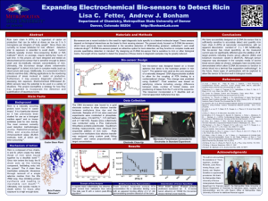
Ricin toxin chain A (RTA), a byproduct of the production of castor oil from castor bean plants, is a hazardous toxin that inhibits the cellular production of proteins once it enters the body. This toxin, whether ingested, inhaled, or injected, can be lethal, and treatment is difficult as there are currently no known antidotes for ricin. Detection of RTA prior to exposure is thus important, and it is necessary to expand the methods that can be used for this detection, as current methods are not time-sensitive. In this project, we have designed and tested an electrochemical biosensor that is capable of– and sensitive enough– to detect small, bio-medically relevant concentrations of RTA. This biosensor was designed based on an existing oligonucleotide aptamers that have been previously shown to bind to the hydrolase protein in ricin toxin. One of these aptamers was then used as the basis of a rationally designed DNA oligonucleotide biosensor scaffold that allows the coupling of RTA binding to a conformational change in the oligonucleotide. Ultimately, this biosensor design allows voltammetric interrogation to detect RTA concentration in complex media (such as coffee, blood, and river water). Additionally, this biosensor is convenient and collects real-time data, offering beneficial applications to the monitoring processes of areas involved in castor oil production. Furthermore, it may possess diagnostic potential in assessing ricin exposure. Electrochemical DNA biosensors have the potential to be used in numerous different situations, and this project shows a strategy for how they may be expanded to the detection and quantification of hazardous toxins.
Derek Clark

Lab Member 2017-2018
Current: Pharmacy Technician
Dr. Ilia Mazin

Lab Member 2016-2017
Current: PhD, the Ohio State University
Dr. Nazar Dubchak
Lab Member 2016-2017
Current: Anesthesiology resident, Walter Reed
Jena Jacobs

Lab Member 2015-2017
Current: DO medical student, A.T. Still University
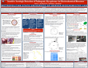
Mycoplasma pneumonia infects 2 million people every year and is responsible for upper respiratory infections and “walking pneumonia.” Here, we describe the creation of a novel electrochemical biosensor capable of detecting pathogenic Mycoplasma for use in academic, research, and clinical applications. Current diagnostics of Mycoplasma, such as molecular-based assays, PCR and serological analysis, are time consuming, expensive, and not particularly accurate. In response, our biosensor is designed for rapid, reliable, and reagentless detection of several common Mycoplasma strains. To do so, we rely on the fact that many pathogenic Mycoplasma share a common secreted protein, P48. A modified aptamer against P48 was incorporated into a custom oligonucleotide scaffold and is used in a gold-electrode-bound fashion to give electrochemical signal change upon binding the secreted P48 target. Ultimately, this biosensor should bring improvements to diagnosis and thus treatment of Mycoplasma in patients who present a proposed infection.
Dr. Jessica (Daniel) Watson
Lab Member 2015-2016
Current: MD, University of Florida
Published!
Susan Jett

Lab Member 2015-2016
Current: Biobank Manager, Thermo Fisher
Aviva Bulow
Lab Member 2014-2016
Current: Software engineer, Sigray
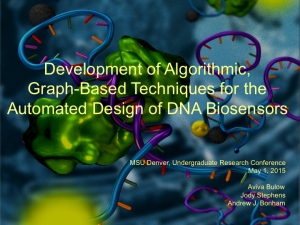
Oligonucleotide-based biosensors have been demonstrated as effective tools for detecting heavy metals, small molecule drugs, and protein targets. Recent efforts to move these biosensors from the lab bench to practical use in industry and medicine rely on the ability to rapidly and effectively adapt them to detect new targets of interest. One compelling aspect of DNA and RNA-based biosensors is the great strides that have been made in computational prediction of the three-dimensional structures that they can assume. However, interpreting predicted structures and using that knowledge to design functional biosensors ab initio remains a challenging problem. We hypothesize that by representing a potential DNA biosensor as a node-weighted graph, we can simplify the challenge of automated structure interpretation and sorting. If correct, this would allow a researcher to provide a core sequence of interest (such as a DNA recognition element or artificially-selected aptamer) and have a software tool design the ideal DNA biosensor for the sensitive detection of that target. This process builds on ideas from our oligionucleotide fold scoring algorithm, Fealden. With this new implementation of Fealden, we are achieving significantly more flexibility, allowing us to analyze any structure type. Because of this we are able to traverse a search space of tens of thousands of possible sequences to determine a “best candidate” biosensor. Additionally, with this increased flexibility we hope to implement some very desirable features in our software, generally, different types of design constraints that a researcher can specify for their biosensor.
Dr. Ryan Warren

Current: Spectrum Health Lakewood, Michigan
Lab Member 2015
Dr. Tiffany (Ashbaugh) Tesmer
 Lab Member 2015-2015
Current: DO, Midwestern University College of Osteopathic Medicine
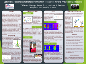
Many modern protein analytical characterization techniques, such as X-ray crystallography, nuclear magnetic resonance, or activity gel shift assays rely on access to large quantities of purified human proteins that have been recombinantly expressed in bacteria. In particular, our lab builds electrochemical and optical biosensors to detect proteins involved in cancer that require pure recombinant proteins for validation purposes. However, the process of expressing and purifying functional human proteins in bacteria requires optimization on a protein-by-protein basis. Here, we investigate varied buffer parameters in the process of affinity column chromatographic separation of recombinant Max (myc-associated factor X) transcription factor protein. The gene encoding a version of Max that is fused to a his-tag purification motif was previously introduced into BL21 E. coli bacteria, and this bacteria were used to produce large quantities of cell lysate. However, the process of isolating Max from the thousands of native bacterial proteins requires careful selection of purification procedures. In particular, the concentration of imidazole, an amino acid mimic used to out-compete Max binding to the affinity column, has dramatic effect on the final purity of the protein obtained. By varying the concentration of imidazole, we aim to allow efficient and optimized purity of Max for testing of our lab’s biosensors.
Michael McCoy

Ebony Miller

Lab Member 2015-2015
Current: Certified Nursing Assistant at University of Colorado Health Sciences Center
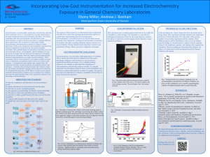
Electrochemistry is an industrially, analytically, and medically important field of chemistry; however, electrochemistry concepts and techniques are rarely introduced in the general chemistry curriculum. Two factors that have contributed to this lack of incorporation are one, that electrochemistry is widely viewed as non-intuitive, requiring additional physics course preparation, and two, because reliable electrochemistry instrumentation has remained outside the budget of student laboratories. In this work, introductory electrochemistry experiments are being investigated for incorporation into the general chemistry curriculum. The guided-inquiry lab experiences will utilize a low-cost electrochemistry device—the CheapStat—that Dr. Bonham et al. have previously developed, allowing us to develop self-guided labs that use instrumentation that can be provided to individual students instead of using limited time on a single classroom machine. These experiments will use electrochemistry to investigate relatable concepts such as verifying the concentration of pain relievers in common cold medicine. It is hypothesized that students who complete these self-guided labs will gain a deeper insight into electrochemical theory and techniques. Currently, labs are being developed and verified to ensure they are reliably reproducible, engaging, and incorporate best practices for self-guided lab experiences. In the future, we plan to assess general chemistry laboratories that complete or do not complete these modules and assess the impact that they make on the educational experience and knowledge of electrochemistry.
Laura Roon

Lab Member 2014-2015
Published!
Jonathan Richards

Lab Member 2013-2015
Published!
Becky Addison

Lab Member 2013-2014
Current: GIS Software Developer at USGS
Jeremy O’Brien

Lab Member 2013-2014
Current: Canning Line Operator at Los Bucaneros
Travis Ingraham

Lab Member 2013-2014
Current: Director of Extraction and Quality Assurance for GCH USA
Sarai Graves

Lab Member 2013-2014
Current: Provider Relations at United Health Group
Kathryn (Norquest) Vang

Lab Member 2012-2014
Current: Masters student, Notre Dame University
Published!
Dr. Stephen Schaffner

Lab Member 2012-2014
Current: DO, Rocky Vista Medical School
Published!
Kyra Brandt

Lab Member 2012-2014
Current: 1st Lieutenant Signal Officer, US Army
Elina (Baravik) Wegner

Lab Member 2012-2014
Current: Oncology R&D at Invitae
Published!
Yerelsy Reyna

Lab Member 2012-2014
Published!
Dr. Josh Sowick

Lab Member 2012-2014
Current: MD, Rosalind Franklin University of Medicine and Science
Jody Stephens

Lab Member 2011-2014
Current: Group Lead Software Testing at ArcherDx
Published!
Dr. Ryan Masterson

Lab Member 2012-2013
Current: Resident doctor, Johns Hopkins
Published!
Mason Preusser

Lab Member 2012-2012
Current: Consultant at D-A Lubricant
Dr. Tonya Santaus

Lab Member 2012-2012
Current: PhD, University of Maryland Baltimore County
Amanda Faux

Matthew Stoddard

Lab Member 2011-2012
Current: Scientific Informatics Consultant at Accenture
Morgan Miller

Lab Member 2011-2012
Current: Digital Strategy Director at DMA Digital